NEWS
January 22, 2014
Spinal cord injuries in young people
Spinal cord injuries often involve young people but few teens and college students understand the potentially life-threatening risks that come with playing many popular sports.
December 23, 2013
Saving Children's Spines
San Diego, California (NAPSI) - While between 1,500 to 2,000 children and adolescents sustain spinal cord injuries every year, you can help keep your kids out of such statistics.
November 20, 2013
Check out this story of inspiration!
Teen determined to walk again after spinal cord injury
A Kansas teenager is walking again after suffering a football injury in September. Watch the inspiring story from KAKE-TV or visit the Team Anthony Crump Facebook page to learn more about his journey.
WHAT IS AN SCI
 Perceptions about the human spinal cord have undergone a revolution in recent years. What was once considered hopeless is now showing signs of promise. SSPF-funded scientists are on the cutting edge of spinal cord research and making progress. Advances are being made every day. This tutorial is designed as an introduction for the layperson to spinal cord injury and repair. It includes information on what happens to the spinal cord as a result of traumatic injury and some of the latest developments in the search for effective treatments.
The central nervous system (CNS) controls most functions of the body and mind. It consists of two parts: the brain and the spinal cord. The brain is the center of our thoughts, the interpreter of our external environment, and the origin of control over body movement. Like a central computer, it interprets information from our eyes (sight), ears (sound), nose (smell), tongue (taste) and skin (touch), as well as from internal organs such as the stomach. It controls all voluntary movement, such as speech and walking, and involuntary movement like blinking and breathing. It is the core of our thoughts, perceptions and emotions.
The central nervous system is better protected than any other system or organ in the body. Its main line of defense is the bones of the skull and spinal column, which create a hard physical barrier to injury. Below the bones is a space filled with cerebrospinal fluid that provides shock absorbance. Unfortunately, this protection can be a double-edged sword. When an injury to the CNS occurs, the soft tissue of the brain and cord swells, causing pressure because of the confined space. The swelling makes the injury worse unless it is rapidly relieved. Fractured bones can lead to further damage and the possibility of infection.
The spinal cord is the highway for communication between the body and the brain. When the spinal cord is injured, the exchange of information between the brain and other parts of the body is disrupted.
Many organs and tissues in the body can recover after injury without intervention. Unfortunately, some cells of the central nervous system are so specialized that they cannot divide and create new cells. As a result, recovery from a brain or spinal cord injury is extremely difficult.
The complexity of the central nervous system makes the formation of the right connections between brain and spinal cord cells very difficult. It is a huge challenge for scientists to recreate the central nervous system as it existed before the injury. Cells called neurons connect with one another to send and receive messages into the brain and spinal cord. Many neurons working together are responsible for every decision made, every emotion or sensation felt, and every action taken. As many as 10,000 different subtypes of neurons have been identified, each specialized to send and receive certain types of information.
Each neuron is made up of a cell body, which houses the nucleus. Axons and dendrites form extensions from the cell body.
Glia cells are "nerve-helper" cells that provide structural support, nourishment and protection for neurons.
Astrocytes, a kind of glial cell, are the primary support cells of the brain and spinal cord. They make and secrete proteins called neurotrophic factors, which assist in the maintenance of nerve cells in the central nervous system. They also break down and remove proteins or chemicals that might be harmful to neurons (for example, glutamate, a neurotransmitter that in excess causes cells to become overexcited and die by a process called excitotoxicity).
Astrocytes aren't always beneficial: After injury, they divide to make new cells that surround the injury site, forming a glial scar that is a barrier to regenerating axons.
Microglia are immune cells for the spinal cord. After injury, they migrate to the site of injury to help clear away dead and dying cells. They can also produce small molecules called cytokines that trigger cells of the immune system to respond to the injury site. This clean-up process is likely to play an important role in recovery of function following a spinal injury.
Oligodendrocytes are glial cells that produce a fatty substance called myelin which wraps around axons in layers. Axon fibers insulated by myelin can carry electrical messages (also called action potentials) at a speed of 100 meters per second, while fibers without myelin can only carry messages at a speed of one meter per second. A spinal cord injury can compromise or destroy myelin.
Messages are passed from neuron to neuron through synapses, small gaps between the cells, with the help of chemicals called neurotransmitters. To transmit a message across a synapse, neurotransmitter molecules are released from one neuron (the "pre-synaptic" neuron) and cross the gap to the next neuron (the "post-synaptic" neuron).
The process continues until the message reaches its destination. There are millions and millions of connections between neurons within the spinal cord alone. These connections are made during development, using positive and negative (inhibitory proteins) signals to fine-tune them. Amazingly, a single axon can form synapses with as many as 1,000 other neurons.
There is a logical and physical topographical organization of the anatomy of the central nervous system, which is an elaborate web of closely connected neural pathways. This ordered relationship means that different segmental levels of the cord control different things and injury to a particular part of the cord will impact neighboring parts of the body.
Paralysis occurs when communication between the brain and spinal cord fails. This can result from injury to neurons in the brain (a stroke), or in the spinal cord. Trauma to the spinal cord affects only the areas below the level of injury. On the other hand, poliomyelitis (a viral infection) or Lou Gehrig's disease (ALS, amyotrophic lateral sclerosis) can affect neurons in the entire spinal cord.
The spinal cord contains multiple tracts to transmit different information. Specialized neurons carry messages from the skin, muscles, joints and internal organs to the spinal cord about pain, temperature, touch, vibration, and proprioception (or spatial orientation). These messages are then relayed to the brain along an ascending pathway, for example the spinothalamic tract. These pathways are in different locations in the spinal cord, so an injury might not affect them in the same way or to the same degree.
Each segment of the spinal cord receives sensory input from a particular region of the body. Scientists have mapped these areas and determined the "receptive" fields for each level of the spinal cord.
Neurons in the motor cortex, the region of the brain that controls voluntary movement, send their axons through the corticospinal tract to connect with motor neurons in the spinal cord. The spinal motor neurons project out of the cord to the correct muscles via the ventral root. These connections control conscious movements like writing and running.
Information also flows in the opposite direction, resulting in involuntary movement. Sensory neurons provide feedback to the brain via the dorsal root. Some of the sensory information is conveyed directly to lower motor neurons before it reaches the brain, resulting in involuntary or reflex movements. The remaining sensory information travels back to the cortex.
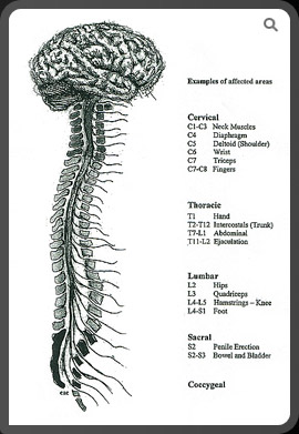
The spinal cord is divided into five sections: the cervical, thoracic, lumbar, sacral, and coccygeal regions. The level of injury determines the extent of paralysis and/or loss of sensation. No two injuries are alike.
In addition to the control of voluntary movement, the central nervous system contains the sympathetic and parasympathetic pathways that control the "fight or flight" response to danger and regulation of bodily functions. These include hormone release, movement of food through the stomach and intestines, and the sensations from and muscular control to all internal organs.
A common set of biological events take place following spinal cord injury:
1. Cells from the immune system migrate to the injury site, causing additional damage to some neurons that survived the initial trauma, and death to others.
2. The death of oligodendrocytes causes axons to lose their myelin, which greatly impairs the conduction of messages or renders the remaining connections useless. The neuronal information highway is further disrupted because many axons are severed, cutting off the lines of communication between the brain and muscles and between the body's sensory systems and the brain.
3. Within several weeks of the initial injury, the area of tissue damage has been cleared away by microglia, and a fluid-filled cavity surrounded by a glilal scar is left behind. Molecules that inhibit regrowth of severed axons are now produced at or near this site. The cavitation is called a syrinx, which acts as a barrier to the reconnection of the two sides of the damaged spinal cord.